Eardrum Perforation
What is eardrum perforation?
An ear drum perforation is a relatively common finding for an otologist (ear specialist). Perforations can come in many different sizes, from small pinholes to complete loss of all eardrum tissue.
What causes eardrum perforations?
The most common cause of perforation is an ear infection. When the infected fluid behind the eardrum builds up, the pressure can become so strong that it bursts open the eardrum and releases out of the ear. The patient usually notices a sudden relief in pressure and pain, but there is drainage from the ear and hearing loss does not improve, even when the infection is cleared.
Other causes of ear drum perforation include:
- Foreign objects (Q-tips)
- Head trauma
- Sudden pressure changes (air travel, skydiving)
- Diving injuries
- Cholesteatoma
- Chronic Eustachian tube dysfunction
- Post-surgical (after ear tube placement)
What are some symptoms of ear drum perforation?
- Hearing loss
- Ear drainage
- Ear pain
- Ringing in the ears
- Dizziness
What tests are required for diagnosis?
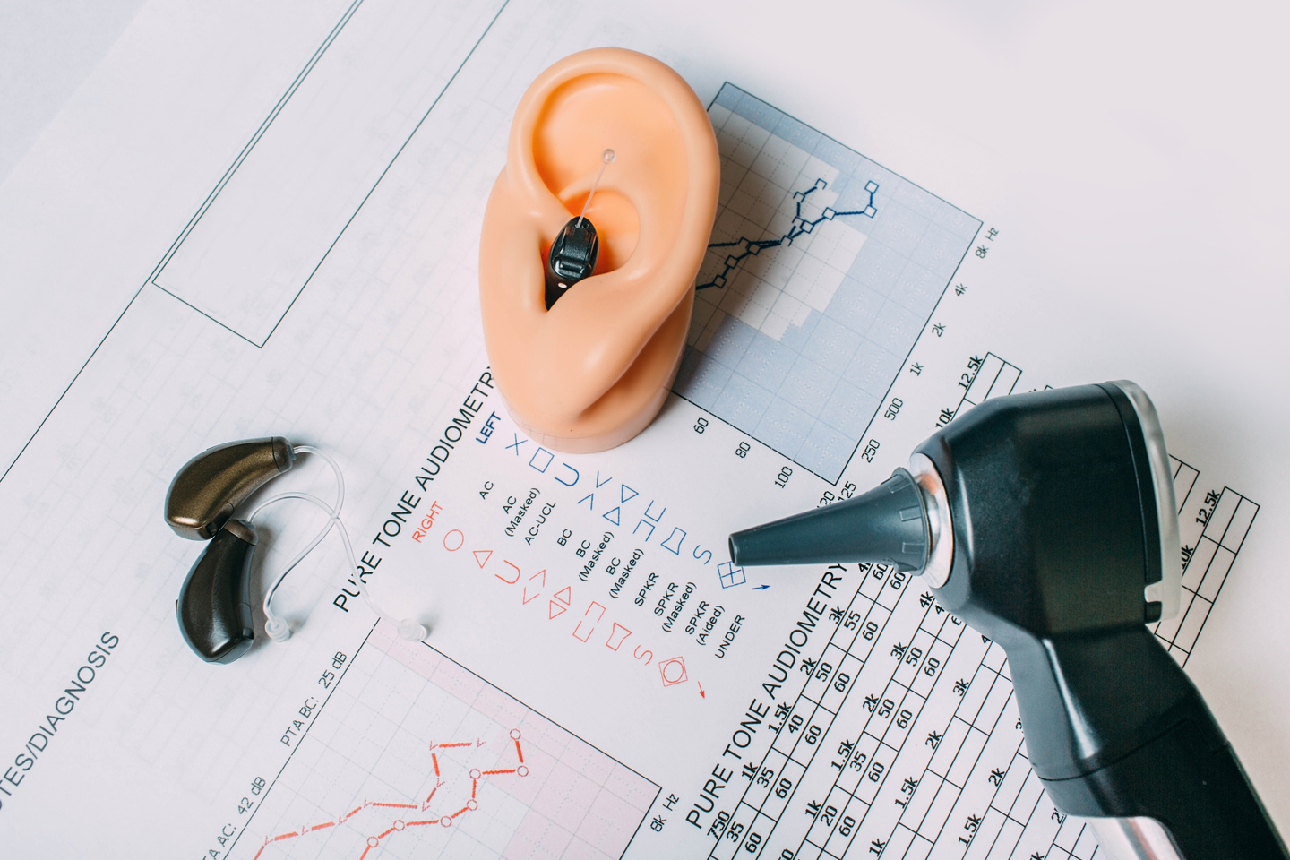
Usually, an otologist (ear specialist) can look in your ear with an otoscope and quickly tell if there is a hole in your ear drum. An otologic microscope is also useful to look through the perforation and assess the condition of the structures behind the eardrum. This helps with surgical planning.
Before a procedure to close the perforation, your otologist may request:
- Audiogram (hearing test)
- Tympanogram (checks movement of the eardrum)
- CT scan (confirms the status of other hidden structures behind the eardrum)
What are my surgical options to repair the hole in the eardrum?
Depending on the size of the perforation, your ear specialist may decide on the best method of treating it.
Small perforations (1-15% of ear drum): These perforations can usually heal on their own after several weeks. If still present, your otologist may offer a paper patch or a fat graft procedure to repair the perforation.
The paper patch procedure is relatively simple and can be performed in the office under local anesthesia. It involves placing a very small and thin piece of paper over the hole, which acts like a band-aid and helps the eardrum grow back appropriately.
Medium perforations (15-35% of ear drum): These perforations are usually too large to close on their own and need some type of surgical intervention. Even though they’re still relatively small, the paper patch may not always be appropriate to successfully close the defect.
Your otologist may offer another minimally invasive procedure known as a fat graft myringoplasty. This procedure can also be performed in the office under local anesthesia. It involves using your own fat to help close the hole. The very small piece of fat is taken from a tiny incision in behind your ear lobe. The fat is then placed inside the perforation and works by promoting your own tissue to heal using the fat as scaffolding material.
Large perforation (35%-100% of ear drum): Large perforations should never be attempted to be repaired with a paper patch procedure. The smaller of these perforations can sometimes be repaired using fat, but usually they require a more formal closure in the operating room. Depending on the size and location of the perforation, your otologist may recommend performing the operation through the ear canal or through a small incision behind the ear. Both operations have their advantages and disadvantages. It is very important that you speak with your otologist about all surgical options.
How do you close a large perforation?
The formal name of the procedure to close a large perforation is called a tympanoplasty.
A tympanoplasty involves making an incision in the ear canal and lifting the skin close to the remaining eardrum. The eardrum is also dissected and elevated, and a graft is placed underneath the perforation to close it. The graft comes from the actual patient: It is usually harvested from a thin layer of tissue taken from the temporalis muscle (the muscle behind the ear) or tragal cartilage (the protrusion in front of your ear canal). The eardrum is then placed back in its original position, and small dissolvable sponges are then placed on top of the newly grafted eardrum to help keep everything in place.
What is the recovery after a tympanoplasty?
The procedure is usually performed in an outpatient surgery center, meaning that patients go home the same day. There is very little pain that can be controlled with simple medications. Patients can usually start doing light activities around the house and office the following day.After 2 weeks, your otologist will examine your ear and remove any remaining packing in the ear. Your hearing may be muffled until the excess packing is removed. Although some patients notice improvement in hearing right after the packing is removed, the majority of patients feel the maximum improvement about 4 to 8 weeks after the surgery.


